Medgital Vision · registered medical device

Holographic surgical navigation

Augmented reality gives the surgeon X-ray vision. CT, MRI and PET data become a hologram aligned to the patient, for diagnostics, planning and navigation right in the operating field.

RZN 2023/21603Registered medical device
7 patentsInventions and utility models
13 programsState software registrations
SkolkovoResident company
500+

surgeries performed in leading Russian clinics

30+

scientific papers and reports since 2020

<5min

to prepare the system for work

150+

anatomical structures segmented automatically by AI

Why it matters

CT and MRI stay outside
the surgical field

Scans give an accurate picture of the patient's anatomy, but during surgery the surgeon can only see them on a monitor.

Long operation time

Searching for and analysing anatomy takes time at every critical step.

Extensive, traumatic access

Complex cases need wide access just to keep the surgical area under control.

Blood loss and complications

Vessels and critical structures stay hidden until the surgeon reaches them.

Attention split with a monitor

To check the images, the surgeon has to look away from the surgical field.

Medgital technology

The X-ray vision effect

With augmented reality the surgeon gains the ability of X-ray vision: every internal anatomical structure that matters is in view, and every action during the operation can be planned from it.
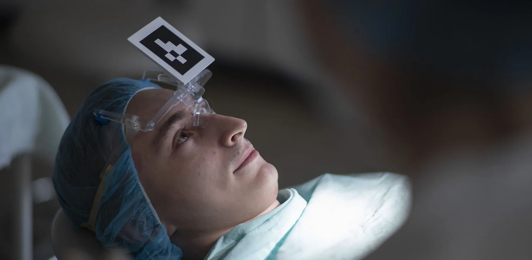
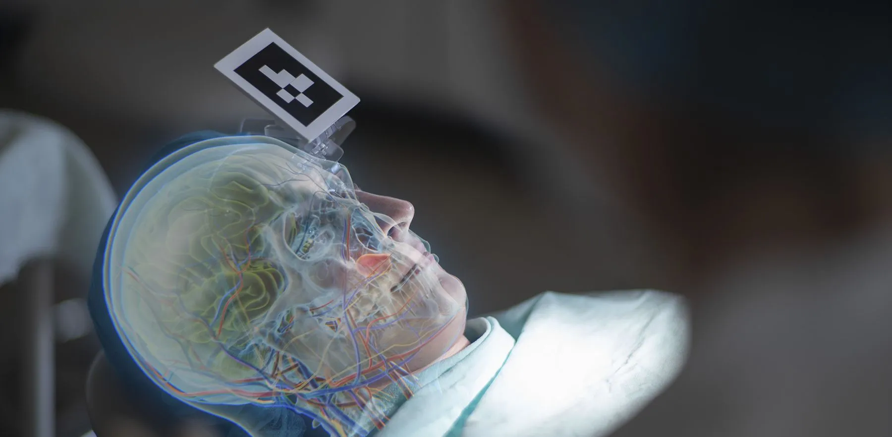
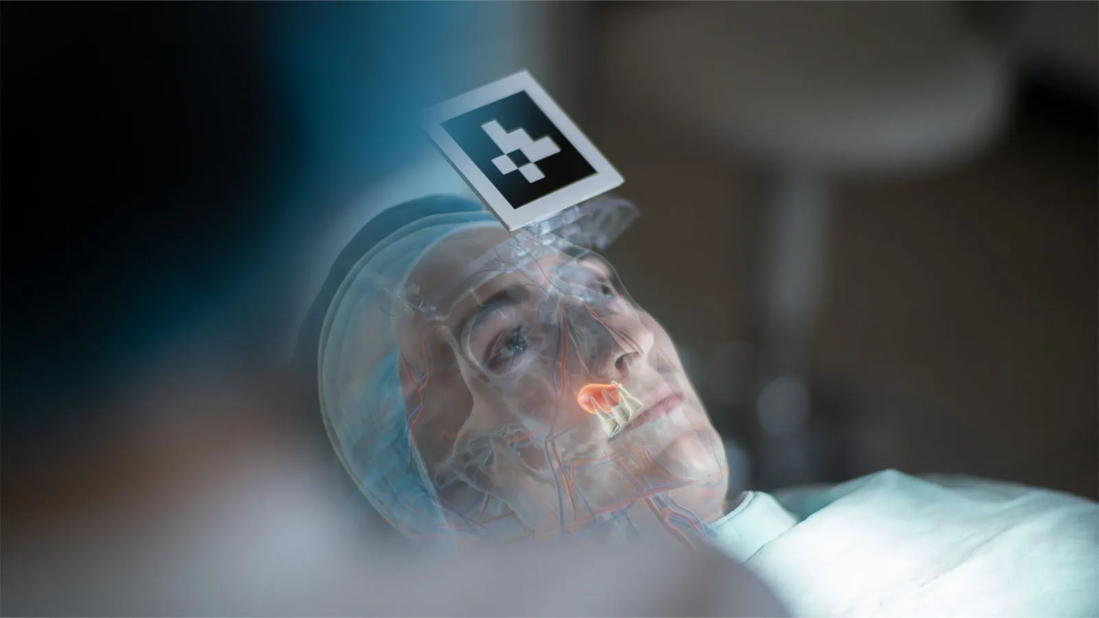
Patient on the operating table with a reference marker on the foreheadThe same patient with a hologram of skull, brain and vessels aligned to the headWith Medgital VisionNaked eye

Precise binding

The hologram is fixed to the patient by Medgital positioning technology, with accuracy under 2 mm.

One volumetric model

Segmented structures and DICOM data are shown together as a single 3D model.

Flexible visualization

The surgeon sees only what is needed at the moment: any structure can be shown, hidden or made transparent.
One workflow

Diagnostics. Planning. Navigation.
In augmented reality.

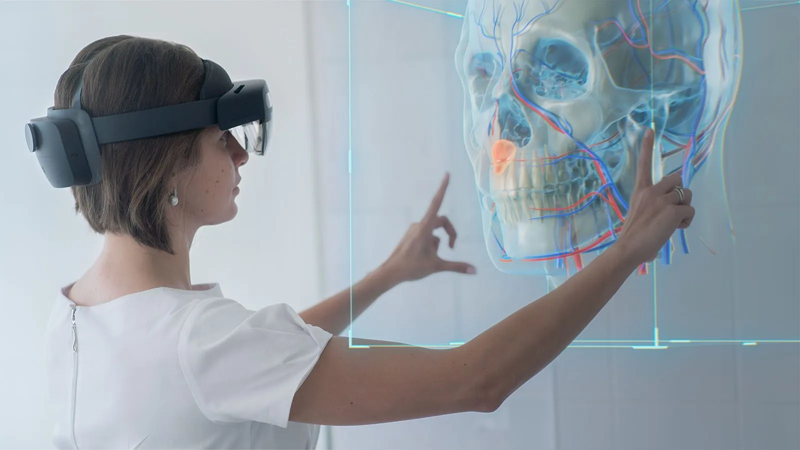
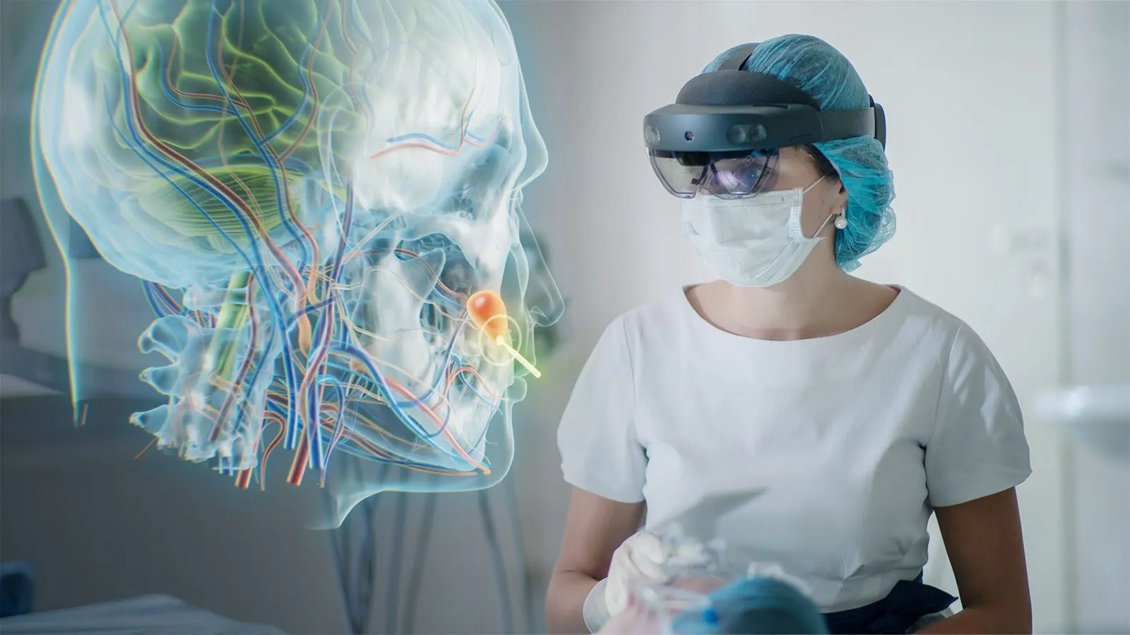
Radiologist in AR glasses examines a holographic skull with vessels

Diagnostics

DICOM data as holograms, right in the radiologist's office.

The patient's CT, MRI or PET study is loaded on the workstation and sent to the AR glasses. The radiologist and the surgeon review the anatomy together, as a hologram, at the patient's side.
  • Transfer to the glasses and a volumetric DICOM model in under a minute
  • AI segmentation of more than 150 anatomical structures
  • CT, MRI and PET fusion in one model
  • Visualization directly on the patient, with sections and segmentation of the areas you need
Surgeon in AR glasses plans an approach next to a holographic head

Planning

Plan the operation without making physical models.

The hologram replaces printed 3D models. The surgical team studies the case at real scale or enlarged, and marks the access directly on the patient.
  • The hologram at real scale or enlarged
  • Surgical access planned on the patient, without soft tissue resection
  • Free 3D sections in any plane
  • Flexible tools for marking the access
Patient with a reference marker and the hologram of the skull aligned to the face

Navigation

Operate without being distracted from the surgical field.

During the operation the hologram stays aligned to the patient. A tracked pointer shows where the instrument is relative to the hidden structures.

  • Everything the surgeon needs is always in view, with no switching to monitors
  • Pointer and instrument tracking with live distances to structures
  • Significant savings in operating room space: the glasses and a compact kit are enough
  • All navigation elements that touch the patient can be sterilized
Medgital Vision software

AI prepares the data.
The surgeon works in the glasses.

Two parts of one workflow: data preparation on the workstation and a holographic interface in the AR glasses.

AI segmentation of a head CT: tumor, ventricles and vessels in a 3D model
Pointer tracking: the instrument and section plane aligned with the hologram

On the workstation

Data preparation

  • Load 3D DICOM dataCT, MRI, PET
  • AI segmentation of anatomy150+ structures
  • Real-time volumetric reconstructionTransfer function
  • Multimodal fusionCT + MRI/PET
  • Import segmentation modelsOBJ, STL
  • Stream the surgeon's view to the screenLive

In the AR glasses

During surgery

  • Move, rotate and scale hologramsGestures
  • 3D sections locked to the pointerReal time
  • Patient registrationLandmarks or markers
  • Patient position trackingReference marker
  • Surgical instrument trackingTracked tools
  • Distances to selected structuresLive
Technology in action

From the skull base to the pelvis

Medgital Vision is used in neurosurgery, maxillofacial, abdominal and ENT surgery. Choose a specialty to see real operations.

External ventricular drain placement: ventricle hologram and tracked pointer
Medgital Vision system

Three configurations,
one software platform

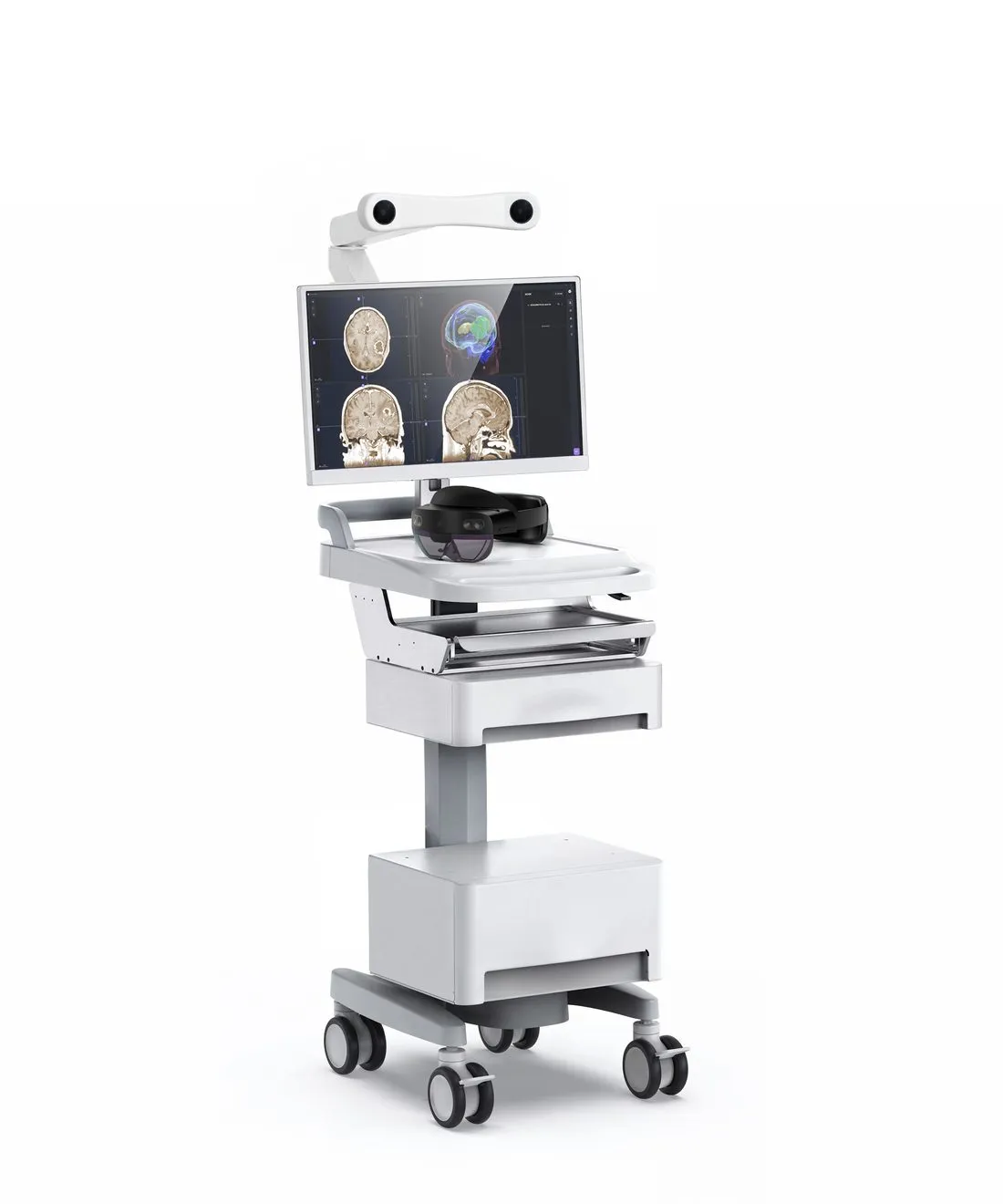
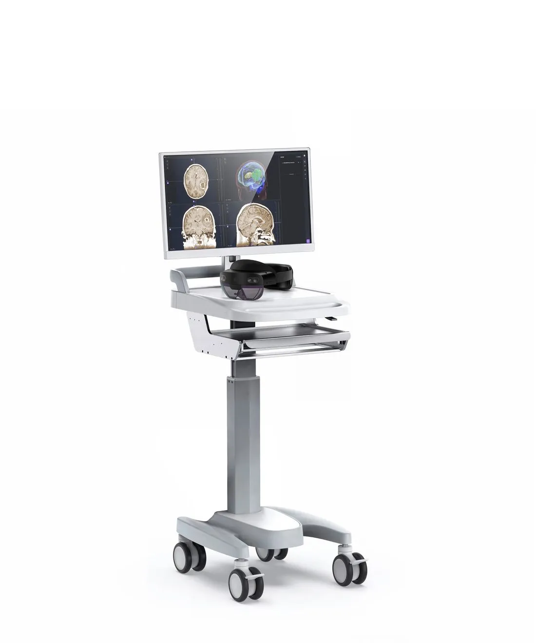
Start with a compact kit for pilot operations and mobile teams, or install a navigation trolley in a high-volume department. Every kit includes the software, AR glasses and the positioning system.
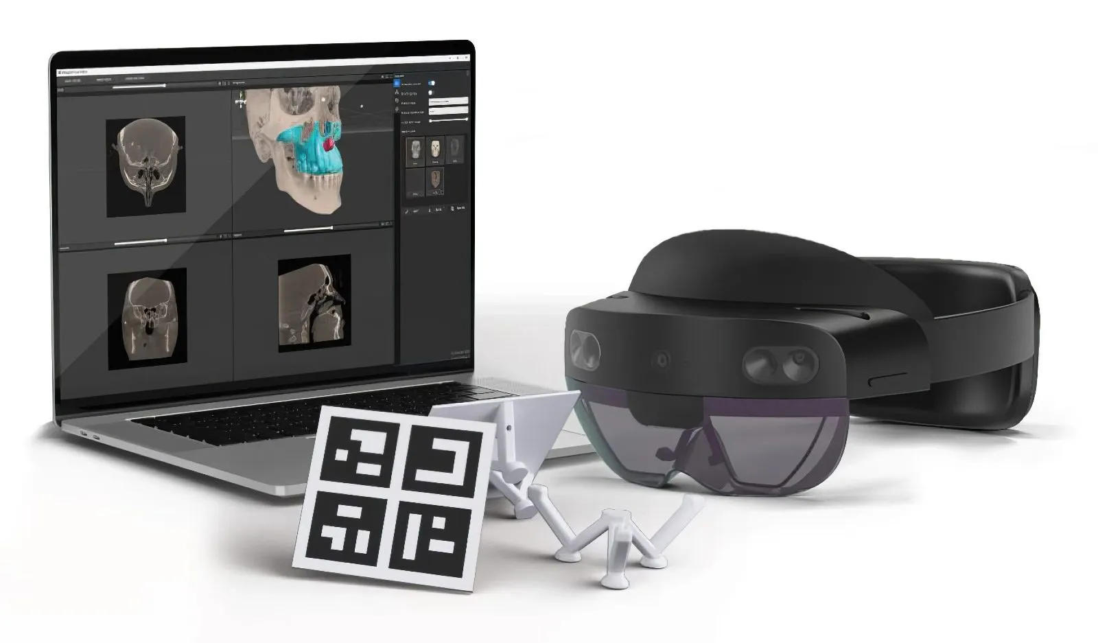
Laptop with Medgital Vision, AR glasses and marker set
Configuration 1

Compact

Fits in a transport case. For pilot operations and mobile teams.
  • High-performance laptop with navigation software
  • Augmented reality glasses
  • Marker system: pointer, reference markers, mounts
  • Wi-Fi router, cables, shockproof case
Medgital navigation trolley with external optical tracking camera
Configuration 3

Trolley + TrackingSoon

External high-precision tracking for complex procedures.

  • Everything in the Trolley configuration
  • External tracking system
  • Additional tracked instruments
  • Expanded markers and mounts
Accuracy< 2 mm
Setup time< 5 min
VisualizationAR glasses
DataCT, MRI, PET
12 months of updatesNew features and fixes
12 months of supportTroubleshooting and service
Staff trainingOnline and on-site sessions
Learning materialsVideo lessons and user manual

Bones, vessels and tumor, aligned to the patient

Pelvic tumor surgery · intraoperative view through the glasses

Reviews

What surgeons and clinics say

Developing a new, original Russian navigation system built on fundamentally different interfaces will enable a breakthrough in modern spinal and cerebral surgery. In routine operations it becomes a necessary layer of patient protection, and it opens the way to exclusive, one-of-a-kind operations.
Prof. D.A. Gulyaev, MD, DScChief neurosurgeon of the North-Western Federal District, Vice-President of the Association of Neurosurgeons of Russia, Professor at the Almazov National Medical Research Centre, Chief Researcher at the Polenov Neurosurgical Institute
Our university is interested in long-term cooperation with Medgital on applying the project's results in maxillofacial surgery and surgical dentistry, and on bringing the system into the clinical practice of our university.
Prof. A.I. Yaremenko, MD, DScVice-Rector for Academic Affairs, Head of the Department of Surgical Dentistry and Maxillofacial Surgery, Pavlov First St. Petersburg State Medical University
The navigation system is of direct practical interest for CT-guided transthoracic biopsy of peripheral lung lesions, including in patients after COVID-19, and for operations on the pelvic organs. We see the project as scientifically and commercially promising in Russia and abroad.
E.V. IvchenkoDeputy Head for Research, S.M. Kirov Military Medical Academy
Evidence

Patents and scientific publications

The technology grew out of research at Peter the Great St. Petersburg Polytechnic University and is described in peer-reviewed journals together with the clinics that use it.

РЗН 2023/21603

Medical device registration certificate

RZN 2023/21603 of 29 November 2023, issued by Roszdravnadzor (Russian Federation). Device: Medgital Vision planning and visualization system, specification TU 32.50.50-001-54095362-2023. Risk class 2a.

7

Patents

Inventions and utility models: marker holders and their fixation for mixed-reality surgery, and navigation for foreign body removal.

13

Software registrations

DICOM reconstruction, CT-to-glasses transfer, surgical navigation, optical instrument tracking, remote visualization control.

Our partners

Clinics, universities and companies
in Russia and abroad

Saint Petersburg

  • Peter the Great St. Petersburg Polytechnic University
  • Pavlov First St. Petersburg State Medical University
  • S.M. Kirov Military Medical Academy
  • Almazov National Medical Research Centre
  • Beloostrov High Technology Clinic
  • St. Petersburg Hospital for War Veterans
  • MVS (Medical Visual Systems)

Moscow

  • Burdenko National Medical Research Center of Neurosurgery
  • Burdenko Main Military Clinical Hospital
  • Sklifosovsky Research Institute for Emergency Medicine
  • National Medical Research Center for Children's Health
  • Kommunarka Moscow Multidisciplinary Clinical Center
  • Russian Center of Neurology and Neurosciences
  • K+31 Clinic
  • VIVET Veterinary Clinic

International partners

  • Intellect X, Minsk, Belarus
  • Bet Medical, India
  • Parul Institute of Medical Sciences and Research, India
  • Instituto Nacional de Cancerología, Mexico

Support

  • Skolkovo Foundation (resident)
  • Russian Science Foundation
  • Foundation for Assistance to Innovations
  • Moscow Innovation Cluster
What's next

AR navigation will become part
of every surgical intervention

A core team of 15 AI and computer-vision engineers is extending the platform in five directions.

Endoscope integration

Endoscopic video combined with holographic anatomy.

Ultrasound navigation

Live ultrasound registered to the 3D model.

Biopsy

Needle trajectory planning and guidance.

Orthopedics

Spine and joint workflows on body-wide segmentation.

Telemedicine

Remote consultation around a shared hologram.

Augmented reality in medicine on Telegram

Clinical cases, new publications and Medgital Vision news on our channel. @ar_in_medicine

Open the channel
Contact us

Learn more about
augmented reality in surgery

Book a demonstration on your own clinical case, or ask us about configurations and delivery. We will prepare an offer for your department.

  • Emailinfo@medgital.org
  • Telegram channel@ar_in_medicine
  • Address6A Lisichanskaya St., premises 14-N, room 2, Saint Petersburg, 197342, Russia
  • CompanyLLC Medgital